In imaging diagnosis, each case is usually made based on an individual radiologist’s memories of past cases with similar imaging features. Sometimes, one may fi nd a variety of imaging fi ndings for a single disease, while, in other cases, different disease entities have similar imag-ing features. As I had once been a trainee in radiology myself, I am fully aware of their diffi cul-ties in dealing with the traditional multiauthor textbooks with drawn-out texts and numerous references. This issue came up during a brief conversation with Dr. Seung Hyup Kim – a uro-radiology expert and my intimate colleague at Seoul National University – several years ago, and Dr. Kim’s sharing of his own experience of writing a new type of textbook provoked me to write this book.
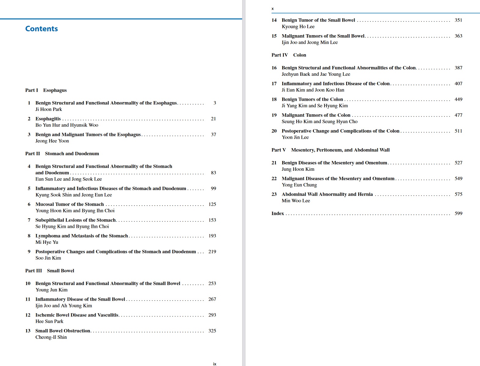
Here, I aim to present a comprehensive textbook of abdominal radiology that would serve as a “visible reference” rather than reading reference for radiology trainees and practicing physicians. The book is designed as a clear, practical, visible guide to the imaging-based diag-nosis of abdominal disease. This volume, the second of two, is devoted to diseases of the esophagus, stomach, duodenum, small bowel, colon, and mesentery, and covers congenital disorders, functional disorders, vascular diseases, benign and malignant tumors, and infl am-
matory conditions. Cases of abdominal wall abnormality, hernia, and postoperative change and complications are also addressed in the volume.
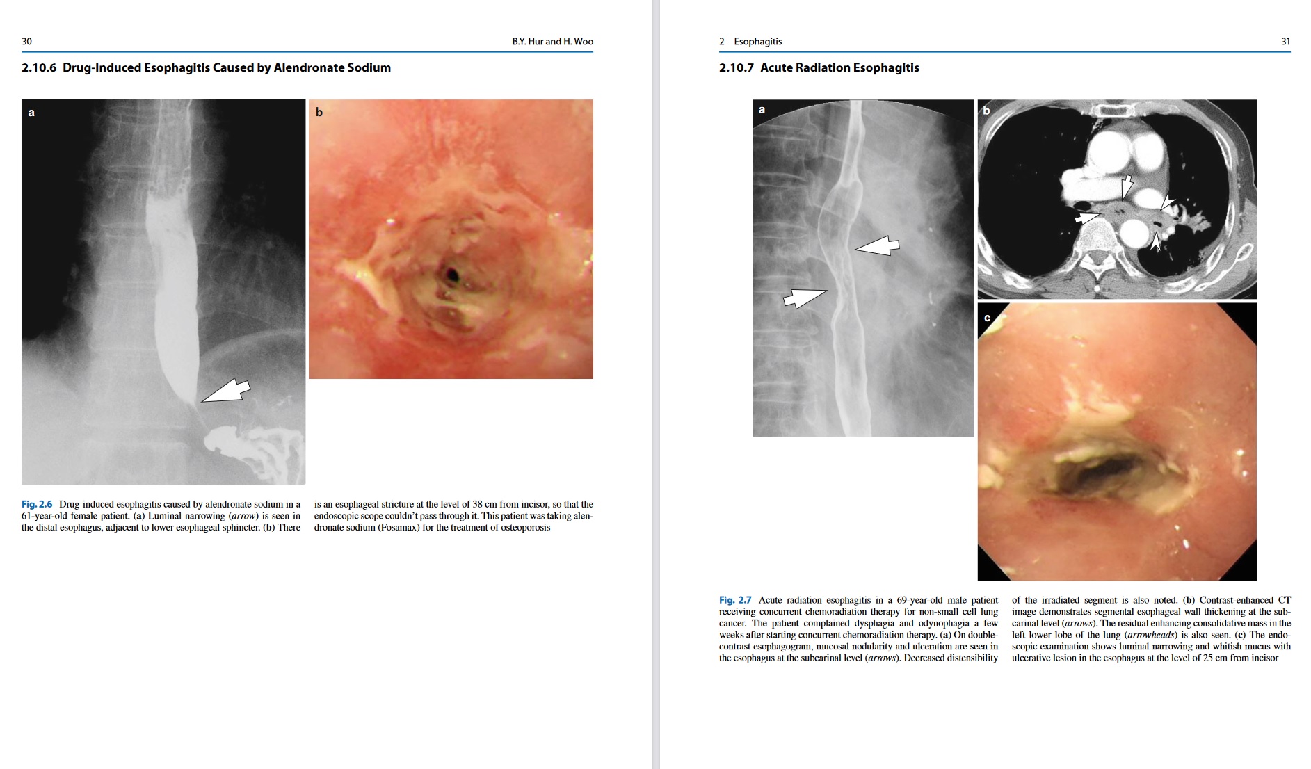
The book presents approximately 400 cases with 1,400 carefully selected and categorized illustrations along with key text messages and tables that will help the reader to easily recall the relevant images as an aid to differential diagnosis. At the end of each text message, key points are summarized to facilitate rapid review and learning. Additionally, brief descriptions of each clinical problem are provided, followed by both common and uncommon case studies that illustrate the role of different imaging modalities such as ultrasound, radiography, CT, and MRI.
I believe that this well-illustrated visible text will be a useful and handy visual guide for radiologists and physicians who interpret diagnostic images of the abdomen in their busy everyday practice.
We are so sorry, but this material is only for VIP members.
PLEASE
SIGN IN
TO DOWNLOAD.
If not VIP member, please
contact us
for more information
